An early predictor of cognitive decline in Parkinson's disease

Have you ever felt the strong sensation that someone is behind you, so intense that you turn around, only to see that no-one is there? This is a 'presence hallucination’. Presence hallucinations are particularly frequent but underreported in patients with Parkinson’s disease and may appear early on in the course of the disease. They are sometimes ignored by the patient, by clinicians, or brushed off as a simple side-effect of medication.

Now, EPFL scientists have found that patients recently diagnosed with Parkinson’s disease and who have early hallucinations are at greater risk of faster cognitive decline. The results are published in Nature Mental Health.

“We now know that early hallucinations are to be taken seriously in Parkinson’s disease,” says Olaf Blanke, Bertarelli Chair in Cognitive Neuroprosthetics, who leads EPFL’s Laboratory of Cognitive Neuroscience.

“If you have Parkinson’s disease and experience hallucinations, even minor ones, then you should share this information with your doctor as soon as possible,” explains Fosco Bernasconi of EPFL’s Laboratory of Cognitive Neuroscience and lead author of the study. “So far, we only have evidence linking cognitive decline and early hallucinations for Parkinson’s disease, but it could also be valid for other neurodegenerative diseases.”

Early hallucinations in Parkinson's disease are associated with a rapid frontal cognitive decline (illustrated by the triangles), and is anticipated by a specific frontal neural oscillation (Theta frequency band).
Early hallucinations in Parkinson's disease are associated with a rapid frontal cognitive decline (illustrated by the triangles), and is anticipated by a specific frontal neural oscillation (Theta frequency band). Credit : EPFL / Bernasconi

A long-term clinical study of Parkinson’s patients

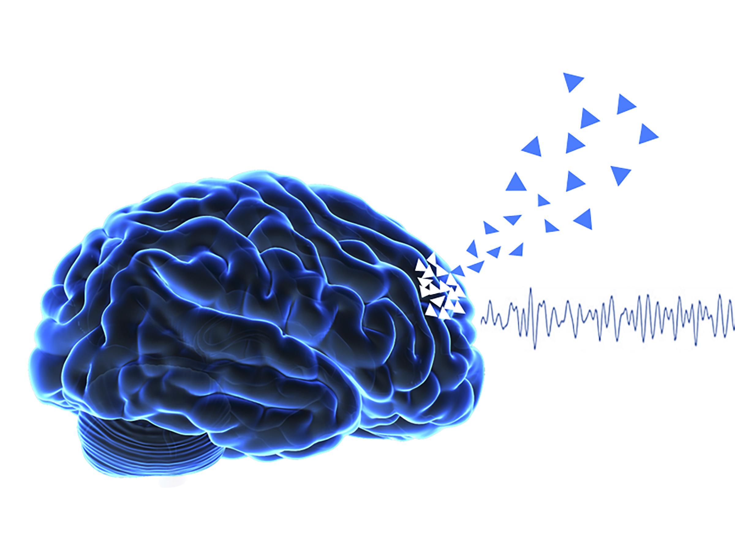
In a collaboration between EPFL and Sant Pau Hospital in Barcelona, the scientists collected data about 75 patients between the ages of 60 and 70 and who were all diagnosed with Parkinson’s disease. The clinicians and scientists at Sant Paolo Hospital conducted a series of neuropsychological interviews to assess their cognitive status, neuropsychiatric interviews about whether or not they were experiencing hallucinations, and electroencephalography (EEG) measurements of the brain’s activity at rest.

In analyzing the data, the scientists found that in patients with Parkinson’s disease, the cognitive decline of frontal executive function is more rapid in the following 5 years for patients with early hallucinations. The level of cognitive decline over those 5 years is further associated with frontal theta (4-8Hz) oscillatory activity as measured by the EEG during the first visit, but only if you have hallucinations at the onset. For clinically and demographically similar patients, the only difference at the outset is that one group has early hallucinations and the other does not.

Early detection for early treatment

Neurodegenerative diseases like Parkinson’s are often detected when it’s too late, the disease too advanced, limiting the impact of preventative measures and disease-modifying therapies. Bernasconi, Blanke and their collaborators are aiming to change that, looking for early signs – like minor hallucinations – and ways to promote early intervention for slowing down progression of cognitive and psychiatric symptoms of the disease.

Hallucinations are among the lesser-known symptoms of Parkinson’s and are highly prevalent early on in the disease, with one individual out of two experiencing hallucinations regularly. Among the different hallucinations, early hallucinations are indeed a cause for concern since they appear in a third of Parkinson patients before onset of motor symptoms like trembling. Parkinson’s disease is traditionally defined as a movement disorder with the typical motor symptoms of resting tremor, rigidity, and bradykinesia, but it also leads to a wide variety of non-motor symptoms that appear early in the course of the disease.

Hallucinations can be described by a continuum of symptoms, from minor symptoms that usually occur early in the course of the disease like presence hallucinations, to more severe symptoms like visual hallucinations that appear later.

It has also already been established that complex visual hallucinations, like seeing someone who is not there, have been linked to cognitive decline and dementia in Parkinson’s disease and related neurodegenerative disorders like dementia with Lewy bodies. However, complex visual hallucinations usually occur at a later stage of the disease, limiting their use as an early marker for cognitive decline.

“Detecting the earliest signs of dementia means early management of the disease, allowing us to develop improved and personalized therapies that try to modify the course of the disease and improve cognitive function,” continues Blanke.

“We aim to have an early marker to identify individuals at risk of a more severe form of Parkinson’s disease, characterized by a more rapid cognitive decline and dementia, based on hallucinations proneness. And ideally identify those individuals even before hallucinations actually occur. We are therefore developing neurotechnology methods and procedures for that purpose,” says Bernasconi.

New real-time guidable-tip wire for surgically treating strokes

Our brains contain an intricate network of arteries that carry blood throughout the organ along winding paths. For neurosurgeons, following these paths with a wire – which is just a third of a millimeter in diameter and enters the body through the femoral artery – to reach an obstructed blood vessel can be tricky. For instance, if they want to point the wire in a different direction, they often have to pull the instrument out and then reinsert it, lengthening surgery times and increasing the risk of complications. But the new wire developed by Artiria is set to change all that. Its tip can be controlled by pressing a button on its handle, through an apparatus that runs entirely on mechanical forces. Artiria just received FDA clearance to test and market its system in the US.

The figures on strokes are startling. According to the World Health Organization, strokes are the leading cause of disability and the second-leading cause of death worldwide. One-fourth of people over 25 can expect to experience one during their lifetime. And when a stroke occurs, time is of the essence – rapid treatment can improve a patient’s prognosis considerably. “While strokes can be caused by a ruptured aneurysm, 80% of the time they’re due to a blood clot in the brain,” says Guillaume Petit-Pierre, Artiria co-founder and CEO. In combination with drug treatments to dissolve the clot, the surgical act, facilitated by the real-time visualization of the instruments by x-rays, makes it possible to extract the clot mechanically. The wire serves as a guide so that the other instruments needed for the operation can be inserted. Before creating their company, Petit-Pierre and Marc Boers – the other Artiria co-founder – spoke with several neurosurgeons and watched them operate several times in order to gain a thorough understanding of the techniques they use. The founders’ goal was to develop a device that would fit in seamlessly with existing procedures. “We were able to get the FDA clearance so quickly because our wire is similar to existing ones in so many respects,” says Petit-Pierre.

These micro-cuts, just a few tens of microns in size, are made from a superplastic alloy, ensuring the necessary flexibility of the wire tip while avoiding injury to the arterioles of the brain.

Guillaume Petit-Pierre

Useful for other types of post-stroke surgeries, too

Petit-Pierre and Boers tested their system on 3D-printed, clear-silicone models of cerebral arteries, and found that it didn’t create any major differences for neurosurgeons. It simply has an extra button on the handle that neurosurgeons can press when they want the tip to bend. A tiny pull wire relays the (slight) mechanical force created from pressing the button all along the structure of the instrument all the way to its 2-centimeter-long deflectable tip. The tip is reinforced on the side connected to the pull wire, and the other side is designed to follow the movement easily. The system may appear simple to the human eye, but fabricating its microscopic-scale parts was a considerable feat of engineering. "These micro-cuts, just a few tens of microns in size, are made from a superplastic alloy, ensuring the necessary flexibility of the wire tip while avoiding injury to the arterioles of the brain. The technological feat also consists in integrating a radio-opaque element into an extremely small volume, enabling the tip of the tool to be visualized during x-ray navigation", explains Guillaume Petit-Pierre. In order to guarantee flawless product cleanliness, the first versions of this system were assembled in EPFL's clean room.

Marc Boers and Guillaume Petit-Pierre © 2023 EPFL

The two founders are also exploring other applications for the underlying technology, which came out of EPFL’s Microsystems Laboratory 4 (LMIS4). “For example, we worked with the Wyss Center in Geneva to see if our wire could be used to lower spasms observed during hemorrhaging storkes,” says Petit-Pierre. Here, the wire could be used to target a specific artery using flexible thin-film electrodes. “There’s currently no effective way to treat cerebral vasospasms, even though they’re known to be a leading cause of disability and death after aneurysm-triggered strokes.”

Petit-Pierre and Boers are old friends and decided to create a startup around ten years ago, while on a backcountry skiing trip. At the time, Petit-Pierre worked in the medtech industry and Boers was already involved in other startups. Petit-Pierre did his PhD at LMIS4 – headed by Philippe Renaud, who was recently named professor emeritus – and the atmosphere there convinced him to try his hand at entrepreneurship. Some 25 businesses have spun off from Prof. Renaud’s lab, so there were plenty of role models to learn from. The core elements of Artiria’s system came from Petit-Pierre’s PhD thesis at LMIS4. With Boers he filed a patent application and created the company in 2019.

Artiria was awarded 2.7 million francs in funding under the European Innovation Council Accelerator Program – although the financing actually came from the Swiss government (SEFRI) since Switzerland no longer has a framework agreement with the EU – and has raised 4.1 million francs from investors. The medtech firm is ranked among Switzerland’s top 100 startups. The two founders plan to launch a more substantial funding round in the coming months, the proceeds of which will be used to expand its seven-person team and validate the product's clinical use.

Four School of Engineering Professors Honored with IEEE Awards

In recognition of their exceptional contributions to their respective fields, four professors from EPFL's School of Engineering have been awarded prestigious honors by the Institute of Electrical and Electronics Engineers (IEEE). The IEEE Awards Program and Best Paper awards are renowned for their acknowledgment of technical professionals who have made a significant impact on technology and society.

Professor Adrian Ionescu, head of the Nanoelectronic Devices Laboratory (Nanolab), has been honored with the 2024 IEEE Cledo Brunetti Award for his groundbreaking work in nanotechnology and technologies for microsystem miniaturization. Ionescu was awarded for his “leadership and contributions to the field of energy-efficient steep slope devices and technologies.”

Professor Andras Kis, head of the Laboratory of Nanoscale Electronics and Structures (LANES), has been granted the 2024 IEEE Lotfi A. Zadeh Award for Emerging Technologies. This accolade celebrates Kis's for his “pioneering work and breakthroughs on 2D materials and electronic devices.” His significant contributions to emerging technologies in recent years have propelled him to the forefront of his field.

Furthermore, Professor Mahsa Shoaran, head of the Integrated Neurotechnologies Laboratory (INL), and Professor Stéphanie Lacour, head of the Laboratory for Soft Bioelectronic Interfaces (LSBI), have jointly received the prestigious 2022 Best Brain Paper (IEEE and SSCS) award. Their winning paper describes a highly scalable and versatile closed-loop neuromodulation system-on-chip (SoC) capable of treating various neurological and psychiatric disorders. Through innovative circuit design techniques and hardware-algorithm co-optimization, Shoaran and Lacour achieved remarkable advancements in channel count, area, and energy efficiency, marking a significant step forward in the field.

The IEEE Awards Program, which has been honoring professionals for nearly a century, recognizes individuals who have made lasting impacts on technology, society, and the engineering profession. The distinguished awards bestowed upon EPFL Professors Ionescu, Kis, Shoaran, and Lacour underscore their outstanding contributions to their respective disciplines and highlight EPFL's commitment to excellence in research and innovation.

Deployable electrodes for minimally invasive craniosurgery

Stephanie Lacour’s specialty is the development of flexible electrodes that adapt to a moving body, providing more reliable connections with the nervous system. Her work is inherently interdisciplinary.

So when a neurosurgeon asked Lacour and her team to come up with minimally invasive electrodes for inserting through a human skull, they came up with an elegant solution that takes full advantage of their expertise in compliant electrodes, and inspired by soft robotics actuation. The results are published in Science Robotics.

The challenge? To insert a large cortical electrode array through a small hole in the skull, deploying the device in a space that measures about 1 mm between the skull and the surface of the brain – without damaging the brain.

“Minimally invasive neurotechnologies are essential approaches to offer efficient, patient-tailored therapies,” says Stéphanie Lacour, professor at EPFL Neuro X Institute. “We needed to design a miniaturized electrode array capable of folding, passing through a small hole in the skull and then deploying in a flat surface resting over the cortex. We then combined concepts from soft bioelectronics and soft robotics.”

From the shape of its spiraled arms, to the deployment of each arm on top of highly sensitive brain tissue, each aspect of this novel, deployable electrode is ingenious engineering.

The first prototype consists of an electrode array that fits through a hole 2 cm in diameter, but when deployed, extends across a surface that’s 4 cm in diameter. It has 6 spiraled-shaped arms, to maximize the surface area of the electrode array, and thus the number of electrodes in contact with the cortex. Straight arms result in uneven electrode distribution and less surface area in contact with the brain.

Somewhat like a spiraled butterfly intricately squeezed inside its cocoon before metamorphosis, the electrode array, complete with its spiraled-arms, is neatly folded up inside a cylindrical tube, i.e. the loader, ready for deployment through the small hole in the skull.

Thanks to an everting actuation mechanism inspired from soft robotics, each spiraled arm is gently deployed one at a time over sensitive brain tissue. “The beauty of the eversion mechanism is that we can deploy an arbitrary size of electrode with a constant and minimal compression on the brain,” says Sukho Song, lead author of the study. “The soft robotics community has been very much interested in this eversion mechanism because it has been bio-inspired. This eversion mechanism can emulate the growth of tree roots, and there are no limitations in terms of how much tree roots can grow.”

The electrode array actually looks like a kind of rubber glove, with flexible electrodes patterned on one side of each spiral-shaped finger. The glove is inverted, or turned inside-out, and folded inside of the cylindrical loader. For deployment, liquid is inserted into each inverted finger, one at a time, turning the inverted finger right side out as it unfolds over the brain.

Song also explored the idea of rolling up the arm of the electrode as a strategy for deployment. But the longer the arm, the thicker it becomes when rolled up. If the rolled-up electrode becomes too thick, then it would inevitably take up too much room between the skull and the brain, placing dangerous amounts of pressure on the brain tissue.

The electrode pattern is produced by evaporation of flexible gold onto very compliant elastomer materials.

So far, the deployable electrode array has been successfully tested in a mini-pig. The soft neurotechnology will now be scaled by Neurosoft Bioelectronics, an EPFL spin-off from the Laboratory for Soft Bioelectronic Interfaces, that will lead its clinical translation. The spin-off was recently granted 2.5 million CHF Swiss Accelerator by Innosuisse.

A neuro-chip to manage brain disorders

Mahsa Shoaran of the Integrated Neurotechnologies Laboratory in the School of Engineering collaborated with Stéphanie Lacour in the Laboratory for Soft Bioelectronic Interfaces to develop NeuralTree: a closed-loop neuromodulation system-on-chip that can detect and alleviate disease symptoms. Thanks to a 256-channel high-resolution sensing array and an energy-efficient machine learning processor, the system can extract and classify a broad set of biomarkers from real patient data and animal models of disease in-vivo, leading to a high degree of accuracy in symptom prediction.

“NeuralTree benefits from the accuracy of a neural network and the hardware efficiency of a decision tree algorithm,” Shoaran says. “It’s the first time we’ve been able to integrate such a complex, yet energy-efficient neural interface for binary classification tasks, such as seizure or tremor detection, as well as multi-class tasks such as finger movement classification for neuroprosthetic applications.”

Their results were presented at the 2022 IEEE International Solid-State Circuits Conference and published in the IEEE Journal of Solid-State Circuits, the flagship journal of the integrated circuits community.

Efficiency, scalability, and versatility

NeuralTree functions by extracting neural biomarkers – patterns of electrical signals known to be associated with certain neurological disorders – from brain waves. It then classifies the signals and indicates whether they herald an impending epileptic seizure or Parkinsonian tremor, for example. If a symptom is detected, a neurostimulator – also located on the chip – is activated, sending an electrical pulse to block it.

Shoaran explains that NeuralTree’s unique design gives the system an unprecedented degree of efficiency and versatility compared to the state-of-the-art. The chip boasts 256 input channels, compared to 32 for previous machine-learning-embedded devices, allowing more high-resolution data to be processed on the implant. The chip’s area-efficient design means that it is also extremely small (3.48mm2), giving it great potential for scalability to more channels. The integration of an ‘energy-aware’ learning algorithm – which penalizes features that consume a lot of power – also makes NeuralTree highly energy efficient.

In addition to these advantages, the system can detect a broader range of symptoms than other devices, which until now have focused primarily on epileptic seizure detection. The chip’s machine learning algorithm was trained on datasets from both epilepsy and Parkinson’s disease patients, and accurately classified pre-recorded neural signals from both categories.

“To the best of our knowledge, this is the first demonstration of Parkinsonian tremor detection with an on-chip classifier,” Shoaran says.

Self-updating algorithms

Shoaran is passionate about making neural interfaces more intelligent to enable more effective disease control, and she is already looking ahead to further innovations.

“Eventually, we can use neural interfaces for many different disorders, and we need algorithmic ideas and advances in chip design to make this happen. This work is very interdisciplinary, and so it also requires collaborating with labs like the Laboratory for Soft Bioelectronic Interfaces, which can develop state-of-the-art neural electrodes, or labs with access to high-quality patient data.”

As a next step, she is interested in enabling on-chip algorithmic updates to keep up with the evolution of neural signals.

“Neural signals change, and so over time the performance of a neural interface will decline. We are always trying to make algorithms more accurate and reliable, and one way to do that would be to enable on-chip updates, or algorithms that can update themselves.”

Locomotion modeling evolves with brain-inspired neural networks

Deep learning has been fueled by artificial neural networks, which stack simple computational elements on top of each other, to create powerful learning systems. Given enough data, these systems can solve challenging tasks like recognize objects, beat human’s at Go and also control robots. “As you can imagine, the architecture of how you stack these elements on top of each other might influence how much data you need to learn and what the ceiling performance is,” says Professor Alexander Mathis at EPFL’s School of Life Sciences.

Working with doctoral students Alberto Chiappa and Alessandro Marin Vargas, the three scientists have developed a new network architecture called DMAP for “Distributed Morphological Attention Policy”. This network architecture incorporates fundamental principles of biological sensorimotor control, making it an interesting tool to study sensorimotor function.

The problem that DMAP is trying to address is that animals – including humans – have evolved to adapt to changes in both their environment and their own bodies. For example, a child can adapt its ability to walk efficiently throughout all the body changes in shape and weight from a toddler to adulthood – and do so on different types of surfaces, etc. When developing DMAP, the team focused on how an animal can learn to walk when its body is subject to these “morphological perturbations” – changes in the length and thickness of body parts.

“Typically, in Reinforcement Learning, so-called fully connected neural networks are used to learn motor skills,” says Mathis. Reinforcement Learning is a machine-learning training method that “rewards” desired behaviors and/or “punishes” undesired ones.

He continues: “Imagine you have some sensors that estimate the state of your body – for example, the angles of your wrist, elbow, shoulder, and so on. This sensor signals are the input to the motor system, and the output are the muscle activations, which generate torques. If one uses fully connected networks, then for instance in the first layer all sensors from across the body are integrated”. In contrast, in biology sensory information is combined in a hierarchical way.”

“We took principles of neuroscience, and we distilled them in a neural network to design a better sensorimotor system,” says Alberto Chiappa. In their paper, published at the 36th Annual Conference on Neural Information Processing Systems (NeurIPS), the researchers present DMAP that “combines independent proprioceptive processing, a distributed policy with individual controllers for each joint, and an attention mechanism, to dynamically gate sensory information from different body parts to different controllers.”

DMAP was able to learn to “walk” with a body subject to morphological perturbations, without receiving any information about the morphological parameters such as the specific limb lengths and widths. Remarkably, DMAP could “walk” as well as a system that had access to those body parameters.

“So we created a Reinforcement Learning system thanks to what we know from anatomy”, says Alberto Chiappa. “After we trained this model, we noticed that it exhibited dynamic gating reminiscent of what happens in the spinal cord, but interestingly this behavior emerged spontaneously.”

Overall, models like DMAP serve two roles: building better artificial intelligence systems based on biological insights, and conversely building better models to understand the brain.

NeurIPS is one of the leading Machine Learning Conferences and many other EPFL labs present their latest work there.

Scientists identify neurons that restore walking after paralysis

In a multi-year research program coordinated by the two directors of .NeuroRestore – Grégoire Courtine, a neuroscience professor at EPFL, and Jocelyne Bloch, a neurosurgeon at Lausanne University Hospital (CHUV) – patients who had been paralyzed by a spinal cord injury and who underwent targeted epidural electrical stimulation of the area that controls leg movement were able to regain some motor function. In a new study by .NeuroRestore scientists, appearing today in Nature, not only was the efficacy of this therapy demonstrated in nine patients, but the improved motor function was shown to last in patients after the neurorehabilitation process was completed and when the electrical stimulation was turned off. This suggested that the nerve fibers used for walking had reorganized. The scientists believe it was crucial to understand exactly how this neuronal reorganization occurs in order to develop more effective treatments and improve the lives of as many patients as possible.

Vsx2 neurons reorganize to restore walking
To arrive at this understanding, the research team first studied the underlying mechanisms in mice. This revealed a surprising property in a family of neurons expressing the Vsx2 gene: while these neurons aren’t necessary for walking in healthy mice, they were essential for the recovery of motor function after spinal cord injury.

This discovery was the culmination of several phases of fundamental research. For the first time, the scientists were able to visualize spinal cord activity of a patient while walking. This led to an unexpected finding: during the spinal-cord stimulation process, neuronal activity actually decreased during walking. The scientists hypothesized that this was because the neuronal activity was selectively directed towards recovering motor function.

To test their hypothesis, the research team developed advanced molecular technology. “We established the first 3D molecular cartography of the spinal cord,” says Courtine. “Our model let us observe the recovery process with enhanced granularity – at the neuron level.” Thanks to their highly precise model, the scientists found that spinal cord stimulation activates Vsx2 neurons and that these neurons become increasingly important as the reorganization process unfolds.

A versatile spinal implant
Stéphanie Lacour, a fellow EPFL professor, helped the research team validate their findings with the epidural implants developed in her lab. Lacour adapted the implants by adding light-emitting diodes that enabled the system to not just stimulate the spinal cord, but also to deactivate the Vsx2 neurons alone through an optogenetic process. When the system was used on mice with a spinal cord injury, the mice stopped walking immediately as a result of the deactivated neurons – but there was no effect on healthy mice. This implies that Vsx2 neurons are both necessary and sufficient for spinal cord stimulation therapies to be effective and lead to neural reorganization.

“It’s essential for neuroscientists to be able to understand the specific role that each neuronal subpopulation plays in a complex activity like walking,” says Bloch. “Our new study, in which nine clinical-trial patients were able to recover some degree of motor function thanks to our implants, is giving us valuable insight into the reorganization process for spinal cord neurons.” Jordan Squair, who focuses on regenerative therapies within .Neurorestore, adds: “This paves the way to more targeted treatments for paralyzed patients. We can now aim to manipulate these neurons to regenerate the spinal cord.”

Scientists decode the neural signals that encode walking in the brain

Deep brain stimulation of the subthalamic nucleus is a well-established neuromodulation therapy for the symptomatic treatment of motor deficits in Parkinson’s disease. For decades, this therapy has been optimized to alleviate symptoms such as tremor, bradykinesia (slowness of movements) and rigidity. However, deep brain stimulation often fails to improve, or can even aggravate gait deficits. To date, little is known about the neural activity patterns underlying gait deficits in Parkinson’s disease, which has restricted the development of neuromodulation therapies better targeting these impairments.

In this study, we leveraged a high-resolution gait platform established at CHUV to record the activity of the subthalamic nuclei, wirelessly and in real time, and to map it to whole-body movements and leg muscle activity while patients performed a series of walking tasks. We identified the neural activity patterns underlying basic walking, turning and freezing of gait. We then developed machine learning algorithms able to predict in real-time different aspects of walking, such as locomotor states, gait phases or effort modulations when avoiding obstacles, as well as pathological episodes such as freezing of gait.

These results open new avenues for the development of adaptive neuromodulation therapies that employ predictions of leg motor states in real time to target and prevent gait and balance deficits in people with Parkinson’s disease.

Link to the article: https://www.science.org/doi/10.1126/scitranslmed.abo1800

Contact information: Eduardo Martin Moraud eduardo.martin-moraud@chuv.ch

About .NeuroRestore
.NeuroRestore is an R&D platform based in French-speaking Switzerland that develops approaches for restoring neurological function in patients suffering from paraplegia, tetraplegia, Parkinson’s disease or the consequences of stroke. It is headed by Grégoire Courtine, a neuroscientist at Ecole polytechnique fédéral de Lausanne (EPFL), and Jocelyne Bloch, a neurosurgeon at Lausanne University Hospital (CHUV). .NeuroRestore, founded in 2018, brings together engineers, doctors and scientists from EPFL, CHUV and the University of Lausanne, with the support of the Defitech Foundation. It draws on this pooled expertise to develop neurotherapies that can help patients recover motor function. Its innovative and personalized treatments are tested through research protocols and then made available to hospitals and patients. .NeuroRestore is also committed to training the next generation of health-care professionals and engineers on the use of these novel therapeutic approaches.

Managing variety in MRI scans can lead to better stroke diagnoses

The first few hours following a stroke are crucial. To be able to treat a patient effectively, doctors must rapidly localize the damaged blood vessel and determine what kind of stroke occurred. In most cases, either a ruptured blood vessel releases blood into the brain, or a blood clot obstructs a blood vessel in the brain. Patients who experience the second type of stroke are prescribed medication to dissolve the blood clot. If this medication is given to patients of the first type, however, it will fluidify the blood and only make the hemorrhaging worse. Yet doctors must take action quickly because the faster a stroke is treated, the lower the likelihood of severe consequences. “Stroke patients are given an MRI [magnetic resonance imaging] scan immediately upon arriving at the hospital,” says Antoine Madrona, who is completing a Master’s degree in life sciences. “The scan is used to confirm that it’s indeed a stroke and to identify what type, in order to prescribe the right treatment.”

Applying AI to medical imaging

Deep-learning algorithms that sort through vast data sets to generate predictions can help doctors make the right diagnosis. They can show radiologists where they should focus their attention, provide quantitative information on the position, size and number of blood vessel injuries, and speed the process of selecting a treatment. As part of his Master’s project, Madrona helped develop an algorithm based on data from diffusion-weighted magnetic resonance imaging (DW-MRI) – a form of MRI that uses the diffusion of water molecules to generate contrast in images. The DW-MRI data sets that Madrona obtained from various Swiss hospitals were extremely heterogeneous. “I was surprised to see so much variation in the images from different hospitals,” he says. “I didn’t expect to see such little standardization.”

In other words, the same patient undergoing an MRI in two different hospitals will end up with two very different images. That’s due to variations in the machine model and the image acquisition protocol. “There’s no national or even international standard,” says Madrona. “Each hospital uses its own magnetic pulse sequence and sets its own pulse directions and intensities. All that affects the image contrast and overall appearance.”

Data security

To make sure patients’ data remain confidential, Madrona used a federated learning method for his algorithm. This method entails training an algorithm across several data sets but without exchanging any data between them. None of the medical imaging systems currently on the market employs a federated method. “I’m particularly interested in how decentralized algorithms can be used to protect patient data,” he says. “This gets to an important ethical issue that I believe needs to be addressed more widely in the healthcare industry. Patient confidentiality shouldn’t be the price to pay for more efficient diagnostics.” Madrona, now 25, intends to pursue a career in this direction.

His Master’s project isn’t quite over, but Madrona already views it as a positive experience. The algorithm could eventually help radiologists in analyzing strokes, regardless of the hospital, MRI machine or image acquisition protocol used. “What really motivates me about my project is the concrete benefits it can deliver directly to clinical applications,” he says.

Another aspect that Madrona appreciates was the opportunity to work with many different institutes. “My project is being supervised jointly by EPFL and the CHUV,” he explains. “I’ve received guidance from Prof. Jean-Philippe Thiran at EPFL’s Signal Processing Laboratory 5 from the School of Engineering, from Jonathan Patiño, a postdoc at the lab, and from Jonas Richiardi, the principal investigator at UNIL’s Translational Machine Learning Laboratory, which is affiliated with the CHUV’s Department of Medical Radiology.” Madrona’s research took place under the umbrella of the Advanced Stroke Analytics Platform (ASAP), a project funded in part by Innosuisse. He also interacted with two other project partners: the Inselspital university hospital in Bern as well as Siemens Healthineers, a pioneer in advanced healthcare technology. A prototype of the new system will be tested at the CHUV and Inselspital in the next six months.

Brain stimulation improves motor skill learning at older age

Even though we don’t think about it, every movement we make in our daily life essentially consists of a sequence of smaller actions in a specific order. The only time we realize this is when we have to learn a new motor skill, like a sport, a musical instrument, a new dance routine or even a new electronic device such as a smart phone or videogame controller.

Perhaps unsurprisingly, there is a lot of research invested in figuring out how humans acquire sequential motor skills, with a majority of studies in healthy young adults. Studies involving older individuals (and common experience) show that the older we get, the harder it is and the longer it takes to learn new motor skills, suggesting an age-related decrease in learning ability.

“As learning is crucial for continuously adapting and staying integrated in daily life, improving these impaired functions will help to maintain the quality of life as we age, especially in view of the constant increase in life expectancy seen worldwide,” says Professor Friedhelm Hummel who holds the Defitech Chair of Clinical Neuroengineering at EPFL’s School of Life Sciences.

In a new study, Hummel and his PhD student Pablo Maceira-Elvira have found that transcranial brain stimulation can improve the age-related impairment in learning new motor skills.

The study used a common way of evaluating how well a person learns new motor skills called the “finger-tapping task”. It involves typing a sequence of numbers as fast and as accurately as possible. The task is popular in studies because it simulates activities that require high dexterity – such as playing the piano or typing on a keyboard – while providing an objective measure of “improvement”, defined as a person increasing their speed without losing accuracy.

Scientists refer to this as a “shift in the speed-accuracy tradeoff”, and it constitutes a key feature of learning. One of the ways the brain achieves this shift is by grouping individual motor actions into so called “motor chunks”: spontaneously emerging brain structures that reduce a person’s mental load, while optimizing the mechanical execution of the motor sequence. “Motor chunks emerge reliably when young adults train on the finger-tapping task, but previous studies show either lacking or deficient motor chunks in older adults,” says Pablo Maceira-Elvira.

Credit: Pablo Maceira-Elvira (EPFL)

The early bird gets the motor skill

The study first trained and tested groups of younger and older adults on learning a new sequence of finger-tapping task, and revealed fundamental differences between the two. Young adults learned the finger-tapping task most efficiently by prioritizing the improvement of the accuracy during their first training session and by focusing on improving their speed thereafter. This led to a shift in the speed-accuracy tradeoff, which allowed efficient motor chunks to emerge early on.

“Older adults showed decreased fast online learning and absent offline learning,” says Maceira-Elvira. “In other words, while young adults show sharp performance increases early in training and improve overnight, older adults improve at a more moderate pace and even worsen overnight.” In contrast, older adults improved their accuracy gradually over the course of training, generating efficient motor chunks only after more extensive practice.

Brain stimulation improvements

Extensive research has been carried out on novel neurotechnologies that may restore learning impairment in older people. “Recent studies have shown we can enhance motor skill acquisition by applying non-invasive brain stimulation to the motor cortex, with anodal transcranial direct current stimulation (atDCS) attracting both academic and commercial interest in recent years due to its unobtrusiveness, portability and affordability,” says Hummel.

In the current study, the researchers applied atDCS to the participants and found that it helped older adults to improve their accuracy sharply earlier on in training and in a pattern similar to that seen in young adults. “Stimulation accelerated the shift in the speed-accuracy tradeoff and enabled an earlier emergence of efficient motor chunks, with 50% of older adults generating these structures during the first training session,” says Maceira-Elvira.

He adds: “The study suggests that atDCS can at least partially restore motor skill acquisition in individuals with diminished learning mechanisms, by facilitating the storage of task-relevant information, quickly reducing mental load and allowing the optimization of the mechanical execution of the sequence.”

“These studies add to the better understanding of age-related deficits in motor skill acquisition and offer a novel strategy to non-invasively restore these deficits,” says Hummel. “These findings open novel opportunities of interventional strategies adjusted to the specific learning phase to restore deficits due to healthy aging or neurological disorder such as stroke.”

Other contributors